Millions of people live with permanent facial redness that forces them to wear foundation daily, makes them uncomfortable in social situations, and which they often believe to be unavoidable. Yet the vascular laser can radically transform this reality — provided the correct diagnosis is made and the right technology is chosen. This article is an informational guide: only a qualified physician can assess your situation and define the appropriate treatment.
1 Redness, a common condition finally treatable
Facial redness, rosacea, telangiectasias and angiomas: these vascular skin conditions affect a significant proportion of the adult population, yet they remain often underdiagnosed and poorly treated. There is frequent confusion between simple reactive flushing — fleeting, triggered by emotion or heat — and a chronic vascular pathology that requires structured medical care.
What is commonly called "couperose" in fact refers to a permanent dilation of the small superficial blood vessels of the skin, visible to the naked eye as red or purplish threads — telangiectasias. Rosacea, on the other hand, is a chronic dermatological disease of which vascular redness is only one of four subtypes. Confusing the two means risking the wrong treatment.
In 2026, the vascular laser is the medical gold standard for treating these conditions. Its principle relies on elegant physics: delivering targeted light energy onto the haemoglobin contained in the dilated vessels, without damaging the surrounding tissues. The result, when the indication is well established and the technology well chosen, can be spectacular — and lasting.
- Facial redness and rosacea are two different entities: one is vascular, the other a chronic multifactorial disease.
- The vascular laser acts on the haemoglobin in vessels without affecting the surrounding skin.
- A precise medical diagnosis is essential before any treatment decision.
- This article is informational only: consult a qualified physician for any therapeutic decision.
2 Understanding redness: diagnosis above all
Before considering laser treatment, it is essential to understand why the vessels have become visible and which underlying pathology is at play. This is not a medical formality — it is the condition for an effective and safe treatment.
The vascular biology of the skin
The skin is traversed by a dense capillary network that ensures its nutrition and thermoregulation. In some people, notably those with fair phototypes (pale, lightly pigmented skin), this vascular wall is naturally more fragile and more reactive. Under the effect of sun, cold, heat or stress, the capillaries dilate abnormally and eventually lose their ability to retract — they then become permanently visible.
The main vascular conditions treated by laser
Facial telangiectasias are fine vessels, isolated or networked, visible on the cheeks, nostrils and chin. They respond excellently to laser. Diffuse facial redness is a generalised form of telangiectasias that gives the complexion a permanent red base. Spider angiomas, also called spider naevi, are small star-shaped lesions particularly frequent in women and during pregnancy. Leg spider veins — telangiectasias and bluish reticular veins — constitute a distinct indication requiring an adapted approach.
Rosacea deserves a special mention: it is a chronic dermatological disease that manifests in four distinct clinical forms. The erythemato-telangiectatic subtype is the most directly treatable by vascular laser. The papulopustular, phymatous and ocular forms require a comprehensive medical management in which the laser is only a complementary tool.
Why prior medical diagnosis is non-negotiable
Some persistent redness is not of benign vascular origin. Lupus erythematosus, dermatomyositis or polycythaemia can present with facial redness — and require specific medical treatment, not a laser. Vascular dermoscopy, performed by a dermatologist, allows precise differentiation of these entities. Treating an undiagnosed redness with laser means risking missing a serious pathology.
- Telangiectasias, diffuse facial redness and spider angiomas are the best indications for vascular laser.
- Rosacea is a chronic disease of which the laser treats only the vascular component.
- Any persistent redness must be examined by a dermatologist before any treatment.
- Vascular dermoscopy is the reference tool for precise diagnosis.
3 How does the vascular laser work?
The vascular laser is based on the principle of selective photothermolysis — a fundamental discovery of the 1980s that revolutionised laser dermatology. The idea is as simple as it is elegant: each structure of the skin has a characteristic chromophore that preferentially absorbs a specific light wavelength. For blood vessels, this chromophore is oxyhaemoglobin — the red pigment of blood that strongly absorbs green and yellow wavelengths.
When a laser pulse is delivered at the right wavelength, the light energy is absorbed by the oxyhaemoglobin and converted into heat inside the vessel. This heat coagulates the vascular wall, causing occlusion of the vessel. The body then naturally eliminates the cellular debris through phagocytosis. The surrounding skin, lacking this chromophore, remains intact.
The precision of this mechanism depends on three fundamental parameters: the wavelength (which determines the targeted chromophore), the fluence (amount of energy delivered) and the pulse duration (which must be shorter than the thermal relaxation time of the vessel to avoid damage to adjacent tissues). It is the mastery of these parameters that distinguishes an expert practitioner from a poorly trained operator.
- The vascular laser targets the oxyhaemoglobin of the blood without affecting the surrounding skin.
- Wavelength, fluence and pulse duration are the three key parameters.
- Mastering these parameters is what differentiates an optimal result from an adverse effect.
- The treated vessel is naturally eliminated by the body within a few weeks.
4 Available technologies: KTP, PDL, Nd:YAG, IPL
Several laser technologies and light sources are available to treat vascular pathologies. Each has its precise indications, advantages and limitations. There is no universally superior laser — the right laser is the one suited to the pathology, the phototype and the location of the lesion.
The KTP laser (532 nm)
The KTP laser (frequency-doubled Nd:YAG) emits at 532 nm, a green wavelength ideally absorbed by oxyhaemoglobin. It excels in treating fine facial telangiectasias, spider angiomas and small superficial vascular lesions on fair skin. Its precision is exceptional, making it the tool of choice for fine, isolated facial vessels. Its main limitation: it is poorly suited to deep vessels or darker skin, where the risk of post-inflammatory hyperpigmentation is higher.
The pulsed dye laser (PDL — 585/595 nm)
Historically the first vascular laser developed, the PDL remains a reference for port-wine stains, haemangiomas and erythematous scars. Its distinctive feature is the transient purpura — a bluish bruise that lasts 7 to 14 days after the session and that reflects vascular coagulation. This purpura, often perceived as inconvenient, has been greatly reduced with modern long-pulse PDLs. Results on port-wine stains and red scars are among the best in all laser dermatology.
The Nd:YAG 1064 nm laser
With its deeper wavelength, the Nd:YAG 1064 nm reaches larger-calibre vessels located deeper in the dermis. It is the only vascular laser truly suited to phototypes IV to VI (darker skin), as it presents a lower risk of hyperpigmentation. It is also used for leg spider veins of small to medium calibre, and bluish facial vessels. The sensation during the session is generally more intense than with the KTP or PDL.
Vascular IPL
IPL (intense pulsed light) is not a laser — it is a polychromatic light source that uses filters to specifically target oxyhaemoglobin. Its major advantage is being able to treat large areas quickly, and to combine vessel and pigment spot treatment in a single session. It is a reasonable option for mild diffuse facial redness on fair skin. Its limitations: less precise and less powerful than a dedicated laser on isolated vessels or complex pathologies such as advanced rosacea.
Comparison table of vascular technologies
| Technology | Wavelength | Main indications | Phototypes | Typical downtime | Effectiveness |
|---|---|---|---|---|---|
| KTP laser | 532 nm (green) | Fine telangiectasias, spider angiomas, facial redness | I to III | Redness 24–48h | Excellent |
| PDL laser | 585–595 nm (yellow) | Port-wine stains, haemangiomas, red scars, rosacea | I to IV | Purpura 7–14 days | Excellent |
| Nd:YAG 1064 laser | 1064 nm (infrared) | Deep vessels, leg spider veins, darker skin | I to VI | Redness 48–72h | Very good |
| Vascular IPL | 515–1200 nm (filtered) | Mild diffuse redness, reactive flushing | I to III | Redness 24h | Good (selected cases) |
- The KTP laser is the reference for fine telangiectasias on fair skin.
- The PDL remains unmatched for port-wine stains and erythematous scars.
- The Nd:YAG 1064 nm is the only vascular laser suited to darker skin.
- IPL is an option for mild facial redness on fair skin, not for complex pathologies.
- The choice of laser depends on the lesion type, phototype and location — not personal preference.
5 Rosacea: a global approach, not just laser
Rosacea is a chronic, inflammatory and multifactorial dermatological disease. The laser can effectively treat its vascular component — but it does not cure rosacea. This distinction is fundamental, and any practitioner presenting the laser as the sole solution to rosacea lacks rigour.
The four recognised subtypes respond differently to laser. The erythemato-telangiectatic subtype — permanent diffuse redness with telangiectasias — is the most directly treatable by KTP, PDL or IPL. The papulopustular subtype — superficially resembling acne — requires medical treatment first (topical metronidazole, azelaic acid, oral doxycycline), with the laser intervening as a complement for the vascular component. The rhinophyma — bulbous skin thickening of the nose — calls for CO2 laser ablation, a distinct surgical approach. Ocular rosacea requires priority ophthalmological referral.
Any vascular laser session must be postponed in case of active inflammatory rosacea flare. Treating inflamed skin increases the risk of adverse effects and may worsen vascular reactivity. Always wait for remission before proceeding.
The triggering factors must be identified and controlled for laser results to be lasting: unprotected sun exposure, alcohol, spices and vasodilating foods, abrupt thermal changes, certain irritating cosmetics. Without this daily discipline, new vessels will appear and results will fade prematurely.
- The vascular laser treats the vascular component of rosacea, not the disease itself.
- Papulopustular forms require medical treatment first.
- Never treat with laser during an active inflammatory flare.
- Managing triggering factors is essential for lasting results.
- Rosacea is managed long-term — it is not definitively cured.
6 Leg spider veins: laser or sclerotherapy?
Leg spider veins — superficial red telangiectasias and bluish reticular veins — are a frequent concern, especially among women. Two medical treatments exist, and their complementarity is often the best approach.
Sclerotherapy consists of injecting a sclerosing agent directly into the vessel, which retracts and is then reabsorbed. It is the reference technique for medium-calibre spider veins and bluish reticular veins. The Nd:YAG 1064 nm laser is preferred for very small red vessels, areas difficult to reach with a needle, or in patients with needle phobia. The two techniques are often combined for optimal results.
It is important to clarify that neither laser nor sclerotherapy treats bulky varicose veins, which fall under vascular surgery or phlebology consultation. A preliminary angiological assessment is recommended to evaluate underlying venous insufficiency before any spider vein treatment.
- Sclerotherapy is the reference for medium-calibre spider veins and reticular veins.
- The Nd:YAG 1064 nm laser is ideal for small superficial red vessels.
- The two approaches are often complementary for optimal results.
- Bulky varicose veins do not fall within laser scope — a vascular consultation is necessary.
7 Choosing your practitioner in Switzerland
In Switzerland, the use of medical lasers is regulated by Swissmedic and governed by cantonal health regulations. Class IV vascular lasers — the most powerful and effective — are reserved for qualified physicians or their delegates under direct medical supervision. Lower-powered IPL devices can be used in institutes, but with significant limitations on the treatable indications.
The dermatologist is the reference practitioner for the diagnosis and laser treatment of vascular skin pathologies. The aesthetic physician trained in laser dermatology can also manage cases of facial redness and telangiectasias. For leg spider veins, the phlebologist or angiologist are the preferred specialists.
The criteria for a serious practitioner are clear: a preliminary medical consultation with clinical examination, a precise diagnosis of the lesion type and phototype, a systematic patch test at the first session, and a personalised protocol. Red flags to avoid: no consultation, treatment based on photos only, non-medical staff operating a class IV laser, promises of guaranteed results.
- In Switzerland, medical vascular lasers are reserved for qualified physicians (Swissmedic, cantonal regulations).
- The dermatologist is the reference practitioner for diagnosis and treatment.
- Require a preliminary medical consultation, a patch test and a personalised protocol.
- Avoid offers without prior consultation or with promises of guaranteed results.
8 A session from start to finish
The initial consultation is the indispensable starting point. It includes a complete skin assessment, vascular dermoscopy to precisely identify the lesion type, determination of the phototype according to the Fitzpatrick scale, and selection of the appropriate laser. A patch test is systematically performed at the first session to verify the skin's response to the chosen laser.
Preparation before each session is simple but non-negotiable: avoid any sun exposure or tanning in the four preceding weeks, do not apply self-tanning cream, and report any current medication — notably anticoagulants and photosensitisers.
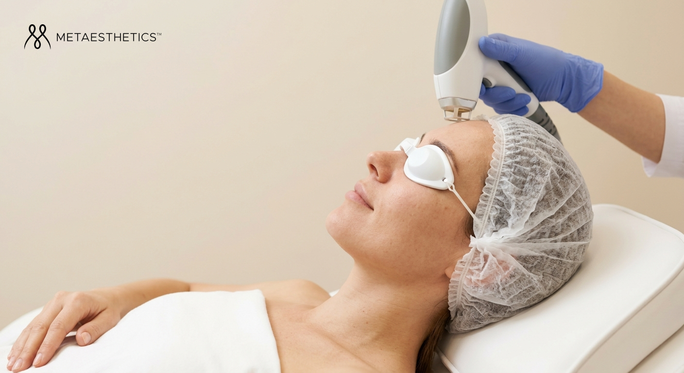
On the day of the session, the area is cleansed and make-up removed, protective eyewear is worn by both patient and operator, and the laser is configured according to the phototype and the type of vessel to be treated. The sensation varies according to the technology: KTP causes light tingling, PDL a rubber-band snap sensation, Nd:YAG a more intense heat. Duration ranges from 15 minutes for localised telangiectasias to one hour for a full face and neck treatment.
The immediate aftermath depends on the laser used. KTP and IPL cause redness and mild oedema that disappear within 24 to 48 hours. PDL produces a purpura (diffuse bluish haematoma) lasting 7 to 14 days — predictable and expected, it reflects the effectiveness of the treatment. In the 48 to 72 hours following the session, avoid sun exposure, intense sport and heat sources. Make-up can generally be resumed the next day for KTP and IPL, after purpura resolution for PDL.
Table: vascular pathology × recommended treatment × prognosis
| Pathology | Recommended laser | Estimated sessions | Prognosis |
|---|---|---|---|
| Fine facial telangiectasias | KTP 532 nm | 1 to 3 | Excellent |
| Diffuse facial redness | KTP, PDL or IPL | 2 to 4 | Very good |
| Rosacea (vascular component) | PDL, KTP | 3 to 6 | Good (long-term management) |
| Spider angioma | KTP or PDL | 1 to 2 | Excellent |
| Port-wine stain | PDL | 5 to 15+ | Variable by extent |
| Leg spider veins | Nd:YAG 1064 nm ± sclerotherapy | 2 to 4 | Very good |
| Darker skin (phototype IV–VI) | Nd:YAG 1064 nm only | 3 to 5 | Good with precautions |
- The initial consultation with patch test is indispensable before the first session.
- Avoid any tanning in the 4 weeks preceding each session.
- PDL purpura is normal and expected — it lasts 7 to 14 days.
- SPF 50+ sun protection is mandatory in the weeks following each session.
9 Results, durability and realistic expectations
The vascular laser delivers often remarkable results — and that is precisely why it is important to set realistic expectations from the first consultation. Isolated telangiectasias and spider angiomas respond excellently, often within one or two sessions: the treated vessels disappear and do not return. Diffuse facial redness generally requires two to four sessions and gives very satisfactory results — with however the possibility that new vessels may progressively appear over time, especially if the triggering factors are not controlled.
Rosacea is the most nuanced pathology in terms of results. The laser significantly improves the vascular component — permanent redness, telangiectasias — but does not alter the chronic nature of the disease. Annual or biannual maintenance sessions are generally necessary to preserve the benefits. Managing triggering factors remains essential.
The durability of results is directly conditioned by a simple but often underestimated element: daily sun protection with a mineral SPF 50+. UV rays stimulate vascular proliferation and inflammation — without rigorous photoprotection, new telangiectasias appear more quickly and results fade prematurely. It is the most profitable investment for preserving the benefits of treatment.
A well-conducted vascular laser can profoundly transform the quality of life of a patient who has been hiding behind foundation for years. But this transformation requires precise diagnosis, an expert practitioner, and daily discipline in sun protection and management of triggering factors.
- Telangiectasias and spider angiomas respond excellently, often in 1 to 2 sessions.
- Rosacea requires long-term follow-up and regular maintenance sessions.
- Treated vessels do not return, but new ones may appear over time.
- Without daily SPF 50+, results fade prematurely.
- The vascular laser truly changes quality of life — provided expectations are realistic.