Pigment laser and dark spots: the complete guide to choose and succeed with your treatment (2026)
Dark spots are one of the most common aesthetic concerns — and one of the most poorly treated. Misdiagnosed, they worsen. Properly managed, with the right laser and the right practitioner, the results can be spectacular and long-lasting. This guide gives you the keys to understand, choose and decide with full knowledge.
Depigmenting creams, peels, "managed" sun exposure: most patients who come in for dark spots have already tried several approaches before considering laser. Some have achieved partial improvement. Others, lacking an accurate diagnosis, have seen their situation worsen. The pigment laser is not a universal solution — it is a powerful medical tool whose effectiveness depends entirely on the accuracy of the diagnosis and the match between the chosen technology and the patient's profile.
1 Dark spots, a common but complex enemy
A dark spot is not simply an excess of colour on the surface. It is the visible sign of a local overproduction of melanin — the skin's natural pigment — by cells called melanocytes. This overproduction can be triggered by the sun, hormones, inflammation, or simply cellular ageing. Understanding the origin of a spot is already half the treatment.
Topical active ingredients — hydroquinone, vitamin C, azelaic acid, retinoids — act by inhibiting melanin production or accelerating cellular renewal. They are effective for prevention and maintenance, but their penetration is limited when pigmentation is anchored deep. This is where laser comes in: by selectively targeting the pigment in the affected layers, it can achieve results that no cream can reach.
Still, you need to choose the right laser, for the right type of spot, on the right phototype. An unsuitable treatment can worsen hyperpigmentation, cause depigmentation or, in the case of melasma, trigger a severe rebound. That is why this article does not merely present the technologies: it emphasises diagnosis as the foundational step of any successful treatment.
- Dark spots result from a local overproduction of melanin with varied causes.
- Topical active ingredients have their limits against deep or established pigmentation.
- The pigment laser is effective but requires an accurate diagnosis beforehand.
- An unsuitable treatment can worsen the situation — especially in the case of melasma.
2 Understanding dark spots: diagnosis first
Not all dark spots look alike, and they are not treated the same way. The first step — non-negotiable — is a medical examination by a dermatologist or a qualified doctor equipped with a dermatoscope, before any treatment decision. This examination makes it possible to identify the exact nature of the lesion, assess its depth within the skin, and rule out a suspicious lesion such as a melanoma.
The main types of dark spots
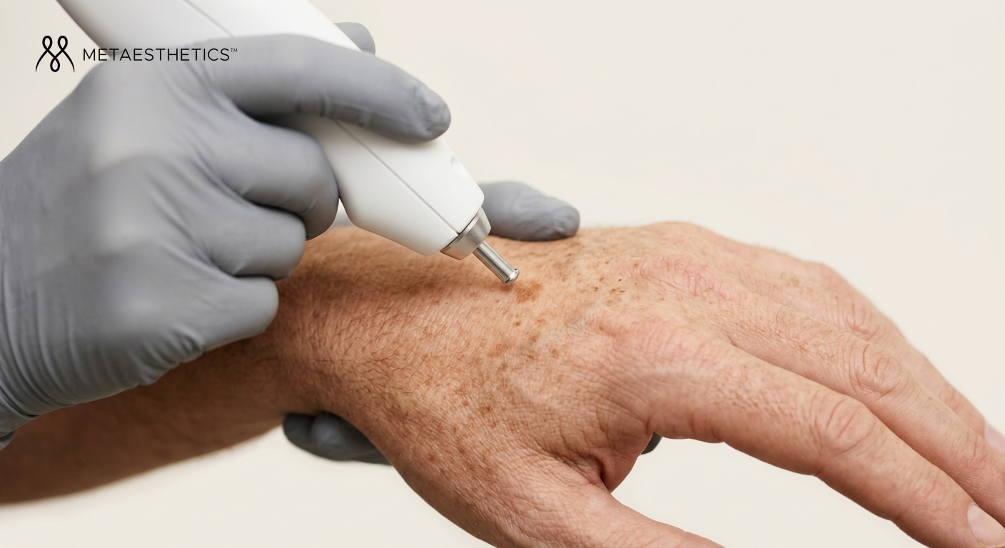
Solar lentigines — often called "age spots" — are the most common. They appear on sun-exposed areas: face, hands, décolleté, forearms. They are epidermal (superficial), well defined, and respond excellently to laser. This is the indication where results are most predictable and long-lasting.
Ephelides — freckles — are genetically determined, fade in winter and reappear in the sun. They can be lightened with laser, but recurrence is inevitable without strict sun protection. Melasma, or the mask of pregnancy, is the most complex form: hormone-dependent, often mixed (epidermal and dermal), and particularly prone to rebound. It deserves a dedicated section in this guide.
Post-inflammatory hyperpigmentation (PIH) occurs after skin trauma — acne, eczema, injury, poorly performed aesthetic procedure. Seborrheic keratoses are benign lesions, often rough, frequently confused with spots but requiring a different laser treatment. Finally, Civatte's poikiloderma affects the neck and décolleté with a characteristic mottled appearance linked to photo-ageing.
Spot depth: a decisive factor
An epidermal spot is located in the superficial layers of the skin. It responds better and faster to laser. A dermal spot is deeper, harder to reach, and the risks of adverse effects are higher. A mixed spot — such as melasma in its most common form — combines both levels and requires a stratified approach. The Wood's lamp and dermoscopy make it possible to assess this depth before any treatment.
- Any examination of a dark spot must begin with a medical consultation including dermoscopy.
- Solar lentigines respond excellently to laser; melasma is far more complex.
- The depth of the spot (epidermal, dermal, mixed) determines the choice of laser.
- The ABCDE rule applies to any suspicious pigmented lesion: consult before treating.
3 How does the pigment laser work?
The pigment laser relies on the principle of selective photothermolysis: a chosen wavelength is preferentially absorbed by melanin, the pigmented target, without damaging the surrounding tissues. The absorbed energy causes thermal or mechanical destruction of the melanosomes — the cellular organelles where melanin is stored — which fragment into microparticles.
These particles are then eliminated by the skin's immune cells (phagocytosis) and gradually cleared. This is why the outcome of a pigment laser treatment is not immediate: it builds over several weeks after the session, at the pace of the natural elimination of the destroyed pigment. On lentigines, this phenomenon is often seen as a transient darkening of the spot, followed by the formation of a thin crust that falls off within a few days — a sign that the treatment has worked well.
The wavelength is the fundamental parameter: each laser emits at a specific wavelength, which determines the depth of penetration into the skin and the selectivity toward melanin. The fluence (energy per cm²) and the pulse duration shape the intensity of the thermal effect. A properly calibrated laser maximises effectiveness while minimising the risk of damaging healthy tissue.
- The laser selectively targets melanin without damaging the surrounding tissues.
- The destroyed pigment is eliminated gradually by phagocytosis — the result appears over several weeks.
- Wavelength, fluence and pulse duration are the key technical parameters.
- Darkening followed by a crust falling off after a session is a normal sign of effective treatment.
4 Available laser technologies
The landscape of pigment lasers has evolved considerably in recent years. Two major families coexist, with complementary indications.
The Q-Switched laser
The Q-Switched laser delivers very short pulses (nanoseconds) at high energy. It exists in several wavelengths: the Nd:YAG 1064 nm penetrates deeper and is suited to dermal spots and olive skin; the Nd:YAG 532 nm (KTP) targets superficial spots on fair skin; the Alexandrite 755 nm is very effective on lentigines and freckles. The Ruby 694 nm completes this arsenal for epidermal pigmentations. It is the reference technology for several decades, with excellent clinical follow-up on solar lentigines.
The picosecond laser
The picosecond laser — whose reference devices include PicoSure, PicoWay and Enlighten — delivers pulses a thousand times shorter than the Q-Switched (picoseconds vs nanoseconds). The effect is more mechanical than thermal: the pigment is fragmented into finer particles, with less residual heat. Recovery is often shorter, and the risk of post-inflammatory hyperpigmentation is lower, making it the technology of choice for complex cases: melasma, PIH, olive skin. In 2026, the picosecond has become the reference for difficult profiles.
IPL and fractional CO2 laser
IPL (intense pulsed light) is not a laser strictly speaking: it emits a broad spectrum, less selective. It can be useful on superficial lentigines of fair skin, but it is strongly inadvisable in cases of melasma — the rebound risk is real. The fractional CO2 laser is ablative: it acts through overall skin remodelling rather than direct pigment targeting. It can be combined with a pigment laser for thick lentigines or seborrheic keratoses, but its use on olive skin requires great caution.
Technology comparison table
| Technology | Wavelength | Targeted spots | Compatible phototypes | Recovery | Efficacy |
|---|---|---|---|---|---|
| Q-Switched Nd:YAG 532 nm | 532 nm | Superficial lentigines, keratoses | I to III | 3–7 days | ✔ Excellent |
| Q-Switched Nd:YAG 1064 nm | 1 064 nm | Dermal spots, olive skin | III to VI | 5–10 days | ✔ Very good |
| Q-Switched Alexandrite | 755 nm | Lentigines, ephelides, freckles | I to III | 3–7 days | ✔ Excellent |
| Picosecond | 532 / 755 / 1 064 nm | Melasma, PIH, complex cases | I to V | 2–5 days | ✔ Reference for difficult cases |
| IPL | Broad spectrum | Superficial lentigines on fair skin | I to III only | 2–4 days | △ Moderate / limited |
| Fractional CO2 | 10 600 nm | Keratoses, thick lentigines, poikiloderma | I to III (caution IV) | 7–14 days | △ Indirect (remodelling) |
Table: spot type × recommended laser × prognosis
| Spot type | Recommended laser | Estimated sessions | Prognosis |
|---|---|---|---|
| Solar lentigo | Q-Switched 532 nm / Alexandrite | 1 to 2 | Excellent |
| Ephelides (freckles) | Alexandrite / Q-Switched 532 nm | 2 to 3 | Good (recurrence possible) |
| Melasma | Picosecond (low fluence) | 4 to 6+ | Complex — long-term management |
| Post-inflammatory hyperpigmentation | Picosecond / Q-Switched 1064 nm | 3 to 6 | Variable depending on depth |
| Seborrheic keratosis | Fractional CO2 / Q-Switched 532 nm | 1 to 2 | Excellent |
| Civatte's poikiloderma | IPL (fair skin) / Q-Switched | 3 to 5 | Good with maintenance |
- The Q-Switched laser remains the reference for solar lentigines on fair skin.
- The picosecond laser is the technology of choice for complex cases (melasma, PIH, olive skin).
- IPL is less selective and contraindicated in cases of melasma.
- The number of sessions varies according to the type of spot, depth and phototype.
5 Melasma: the most complex case
Melasma deserves special attention, as its management differs so much from other hyperpigmentations. It is a hormone-dependent condition — often triggered by pregnancy, oral contraception or the sun — that preferentially affects women with medium to high phototypes. It appears as symmetrical brown patches on the face, particularly the forehead, cheeks and upper lip.
Its complexity lies in its mixed nature: pigment is often present in both the epidermis and dermis, and the melanocytes involved are hyperreactive. Any source of heat or inflammation — including a poorly calibrated laser — can trigger a rebound phenomenon, where the skin produces even more melanin in response to the aggression. This is why melasma has long been considered a relative contraindication to laser.
Current protocols have evolved. In 2026, the low-fluence picosecond laser and LADD protocols (Low Fluence Q-Switched) are the consensus for melasma: they allow action on the pigment without triggering the inflammatory reaction that promotes rebound. These approaches are always combined with a topical treatment (tranexamic acid, mild depigmenting agents) and an absolutely strict SPF 50+ sun protection. Without the latter, no laser treatment of melasma holds over time.
It is essential to be honest with patients who suffer from melasma: it cannot be cured definitively. It is managed over the long term, with regular maintenance protocols, permanent sun vigilance and therapeutic adaptation to hormonal fluctuations.
- Melasma is hormone-dependent, often mixed (epidermal + dermal) and prone to rebound.
- A poorly calibrated laser can worsen melasma — the choice of technology is critical.
- Low-fluence picosecond is the recommended protocol in 2026.
- Melasma is managed over time — it cannot be cured definitively.
- SPF 50+ sun protection is non-negotiable to maintain results.
6 Phototypes and spots: tailoring the treatment to each skin
The phototype — defined by the Fitzpatrick scale in six levels, from the fairest (I) to the darkest (VI) — is the first parameter assessed before any pigment laser treatment. It determines the level of baseline melanin in the skin, and therefore the risk of confusion between the therapeutic target (the spot) and the pigmented background of the skin itself.
Phototypes I to III (fair to slightly olive skin) are the best candidates for pigment laser. The contrast between the spot and healthy skin is maximal, laser selectivity is optimal, and the risks of adverse effects are minimised. Results on solar lentigines are often spectacular from the first session.
Phototypes IV to V (olive to dark skin) can be treated, but with increased caution. The main risk is post-inflammatory hyperpigmentation: the skin reacts to the laser aggression by producing more melanin, which can create a spot more visible than the one that was being treated. The picosecond laser, reduced fluences and a prior depigmenting preparation (4 to 6 weeks) make it possible to minimise this risk. Phototype VI is a very restrictive indication, reserved for very specific protocols with rigorous medical follow-up.
- Phototype is the first assessment criterion before any pigment laser treatment.
- Phototypes I to III obtain the best results with the fewest risks.
- Phototypes IV to V require depigmenting preparation and a suitable protocol.
- Phototype VI is a very restrictive indication — only a highly experienced practitioner can treat it.
7 Choosing your practitioner in Switzerland
In Switzerland, the use of medical lasers for aesthetic purposes is regulated by Swissmedic and cantonal regulations on medical practice. Class IV pigment lasers are medical devices whose use is reserved for doctors or qualified healthcare professionals under medical supervision. Regulations vary from canton to canton: it is therefore important to check your practitioner's credentials.
The ideal practitioner for a dark spot treatment is a dermatologist or a doctor specialising in aesthetic medicine, specifically trained in laser therapy and equipped with several technologies in their practice. The initial consultation should systematically include a complete skin assessment, dermoscopy of the lesions, determination of the phototype and a patch test if necessary.
Certain signals should alert you: no prior medical consultation, treatment proposed without examination of the lesion, non-medical staff handling a Class IV laser, or the promise of guaranteed results in a single session regardless of the nature of the spot. Medical tourism for pigment laser presents specific risks: in case of complication (rebound, depigmentation), remote follow-up is difficult, even impossible.
- In Switzerland, medical lasers are regulated by Swissmedic and cantonal regulations.
- Prefer a dermatologist or aesthetic doctor trained in laser therapy.
- The consultation should include dermoscopy, phototype assessment and patch test.
- Lack of prior assessment and promises of guaranteed results are warning signs.
8 Procedure, results and risks
How a session unfolds
The session begins with cleansing of the area to be treated, followed by the placement of appropriate eye protection. The practitioner sets the laser parameters according to the type of spot, phototype and area. A patch test on a small area may be performed during the first session. The sensation varies by technology: intense heat, slight pricks, or even a sharp snap with the Q-Switched. The duration ranges from fifteen minutes for a small area to one hour for an extensive treatment of the face and hands.
After the session, immediate aftereffects include redness, slight swelling and, on lentigines, a characteristic darkening of the spot. In the following days, a thin crust forms and falls off spontaneously — it is imperative not to pick at it. Make-up can generally be resumed after 5 to 10 days depending on the laser used. SPF 50+ sun protection is mandatory from the day after and for the following weeks.
Results and durability
Solar lentigines respond spectacularly to laser, often from the first session. Durability is excellent if sun protection is maintained. Ephelides fade well but recur with sun exposure. PIH responds variably depending on depth and phototype. Melasma improves with the right protocols, but maintenance sessions are needed over the long term. Spot reduction is often 70 to 90% after a complete course depending on the types of spots.
Risks and contraindications
Within a rigorous medical framework, the pigment laser has a favourable safety profile. The most frequent adverse effects are transient: redness, sensitivity, crusts. Post-inflammatory hyperpigmentation is the main risk, especially on high phototypes — it is largely predictable and avoidable with good preparation. Depigmentation (hypopigmentation) is rare but possible, particularly in cases of excessive fluence. Melasma rebound is a real risk if the technology or fluence is unsuitable.
Absolute contraindications include: recent sun exposure or active tan, pregnancy, use of photosensitising medications (certain antibiotics, oral retinoids, amiodarone), and any undiagnosed suspicious pigmented lesion. Relative contraindications include a history of keloids, a recent shingles outbreak on the area to be treated, and certain active skin diseases.
- Darkening followed by a crust falling off on lentigines is a normal sign of effective treatment.
- Never pick at crusts — risk of scarring and PIH.
- SPF 50+ is mandatory after any pigment laser session.
- PIH is the main risk on olive skin — largely predictable with good preparation.
- Active tanning and pregnancy are absolute contraindications.
9 Complementary treatments and prevention
The pigment laser does not act alone in a well-conducted protocol. Depigmenting chemical peels — with glycolic, kojic or tranexamic acid — are excellent maintenance treatments between laser sessions, or an alternative for profiles who cannot benefit from laser. Microneedling, combined with depigmenting active ingredients, can be useful as a complement on mild PIH, but must be used with caution on olive skin.
Topical active ingredients play a fundamental role before and after laser. Oral or topical tranexamic acid is particularly promising in melasma. Vitamin C and retinoids, used for maintenance, slow melanogenesis and accelerate cellular renewal. These active ingredients do not replace laser on established spots, but they prolong its results.
Prevention remains the most rewarding investment. Daily photoprotection with SPF 50+ — applied correctly, renewed every two hours in case of exposure — is the only measure that sustainably preserves the results of a laser treatment and slows the appearance of new spots. Dietary antioxidants (polyphenols, vitamin C, omega-3) and stress management (cortisol stimulates melanogenesis) complete this preventive approach. No laser treatment, however effective, holds without this daily discipline.
A successful treatment is always a personalised one: the right laser, on the right type of spot, for the right phototype, with the right follow-up. The skin has its own rules — respecting them is the condition for any lasting result.
- Depigmenting peels and topical active ingredients complement and prolong the effect of laser.
- Tranexamic acid is particularly useful in the management of melasma.
- Daily SPF 50+ is the sine qua non condition for the durability of results.
- Prevention (photoprotection, antioxidants, healthy lifestyle) is worth as much as the treatment.